
Using Infared Photography
in Dermatology
Authors: Eliza Hutchison, Humaira Ahmed, Saumya Singh
Chief Editor: Dr Daniel Keith
Infrared (IR) photography is long established as an art form, allowing images to be created using light outside the visible spectrum. Professional and amateur photographers have developed several ways of seeing the world through this hidden light. These include IR film, filters and specialized cameras. One method is converting an old digital camera to IR by removing its internal IR filter from the existing sensor, result- ing in a cheap but effective way of capturing IR photographs. The use of infrared photography in medicine has been documented in forensic pathology, where it helps document post mortem findings, including gunpowder traces, victim identification via tattoos, and detection of external signs of blunt force trauma such as bruises.
In living patients, differentiating between deep dermal pigmentation, such as lumbosacral dermal melanocytosis, and contusions from nonaccidental injury has proven challenging in the busy environment of the emergency department.
The following study aimed to find out whether this IR photographic technique could be an easy way to rapidly tell the difference.
Photographs from a series of patients with either bruising or haematomas as a result of trauma or deep dermal pigmentation using conventional visible light photography and an IR-converted digital SLR camera (IR-converted Nikon D50 with Nikon AF 35–80 mm F/4–5.6 lens) with a mounted IR torch. All IR photographs were colour corrected using open-source image editing software.
Enhanced visualisation of bruising and blood-containing structures through the sharper contrast under IR photography was demonstrated. In comparison, melanin sources of pigmentation, such as deep dermal pigmentation, were less obvious if not invisible when photographed in IR.
Examples included photographs of lumbosacral dermal melanocytosis and oculodermal melanocytosis (naevus of Ota).
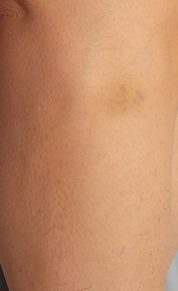
A bruise

Naevus of Ota
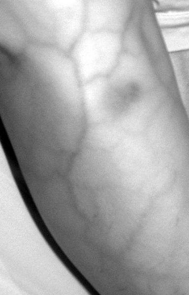
The same bruise becoming more obvious in infrared light

The same naevus of Ota becoming less obvious in infared light
Therefore, IR photography using a modified digital camera may be a simple, cheap and effective technique allowing the rapid and noninvasive identification of skin bruising, differentiating it from deep dermal pigmentation. This is particularly important in the setting of the emergency department, where confusion has occurred, particularly in patients with skin of colour.
Humaira Ahmed, Eliza Hutchison, Saumya Singh, Hagar Elgezeri, Fangyi Xie, Charankumal Singh Thandi, Daniel Keith, PA22 A simple budget-friendly technique in infrared skin photography for the rapid differentiation of deep dermal pigmentation from bruising, British Journal of Dermatology, Volume 193, Issue Supplement_1, July 2025, ljaf085.381, https://doi.org/10.1093/bjd/ljaf085.381
Instructions to set up your camera for infrared skin photography for the rapid differentiation of deep dermal pigmentation from bruising
Cameras primarily use visible light to take photographs. However, infrared photography is a long-established art form used to create unique photographs. It has also been used in forensics to document post-mortem findings, including external signs of blunt force trauma such as bruises.
The use of infrared photography is not yet routine in other areas of medicine. However, it can be used to help differentiate between deep dermal pigmentation such as lumbosacral dermal melanocytosis (also known as Mongolion blue spot), and contusions from non-accidental injury. This can be a challenging but essential distinction for clinicians to make.
Here we outline a budget-friendly, step-by-step guide to set up your own modified digital camera to take infrared photographs for this purpose.
The Camera
Any digital camera can be converted into an infrared camera. You can either do this yourself or pay for an inexpensive professional service to do this for you.
An example set of instructions can be found here.
• [Get great results by converting an older digital camera to infrared | Amateur Photographer]
• [How to turn your DSLR into a full spectrum 'super camera' | Extremetech]
• Infrared Conversion: How To Convert A Camera To Infrared
Dermoscopea takes no responsibility for modifications made to your camera. Please take care and be aware once modified the camera cannot be or is very difficult to restore to how it was. For this reason we recommend using a camera you no longer need and wouldn’t care if it got broken.
The best camera to do this with is a Digital Single Lens Reflex (D-SLR) camera or Mirrorless Interchangeable Lens camera, though it is possible with any digital camera. For the purpose of this article we converted an old Nikon D50 from 2005.
Instructions

1) Lay out your equipment.
You will need:
a) A digital camera (here we use a infrared converted Nikon D50 DSLR Camera with Nikon AF Nikkor 35-80mm lens)
b) An infrared torch (can be purchased from Ebay for around £5) (Optional – This can be skipped if able to take photographs outside in natural daylight)
2) Attach the lens to the lens mount on the front of the camera


3) Attach the infrared torch to the flash mount
4) Ensure the mode dial is switched to ‘P’ (programmed auto)


5) Ensure the IR torch is switched to half mode
6) Take your photograph


7) Colour-correct your images using GIMP (GNU Image Manipulation Program), open-source software available free to download from https://www.gimp.org
Modifications are optional. However, they make the images easier to interpret and can be accomplished with free to use software.
We used GIMP v3.0.4 for GNU/Linux running in Xubuntu 24.04.
Recommended edits are simple and automated
• Convert to Grayscale
• Auto White Balance
Steps complete! You now have your infrared skin photograph which will highlight vascular features while suppressing melanin signals.