
Author: Dr Elina Pantelidi
Chief Editor: Dr Daniel Keith
Rosacea is a long-standing inflammatory condition of the skin. It is estimated to affect 1%-
22% of the general population.
The exact pathophysiology is not known and is thought to be multifactorial. Contributing
factors include genetic predisposition, inflammation and neurovascular mechanisms.
Potential triggers include UV exposure, alcohol, inflammation and others.
Typical presentation:
- Redness/erythema
- Telangiectasia
- Papules
- Pustules
As seen in the pictures below, rosacea usually presents with centrofacial involvement.


Rosacea affecting the nose, cheeks, chin and forehead
Subtypes:
-
Erythematotelangiectatic
-
Papulopustular
-
Phymatous
-
Granulomatous (specific variant of rosacea)
-
Lupus miliaris disseminatus faciei (LMDF)
-
Rosacea-like demodicosis
Even though different subtypes of rosacea have been identified, it is important to note that there is overlap between the different types.
Dermoscopic Appearance
Dermoscopy can be utilised to aid in the diagnosis of rosacea.
The main finding of dermoscopy is the presence of polygonal vessels, usually in a reticular arrangement, with a regular or patchy distribution.
Other findings include follicular plugs, follicular pustules, yellow clods, demodex tails and white amorphous areas.
Contrary to the other types of rosacea, in phymatous rosacea, the main dermoscopic findings are follicular findings. Vascular findings are less evident.


Linear reticular vessels – reticular polygonal pattern

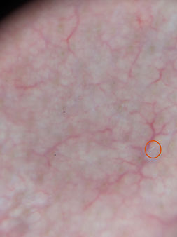
Reticular polygonal vessels
Red circle: Demodex tail


Linear reticular vessels, regular polygonal pattern
Blue star: white amorphous areas
White arrow: demodex tail
Red circles: follicular plugs
Green star: red diffuse structureless area


Phymatous rosacea
Polymoprhic/non specific vessel morphology
Red arrows: follicular yellow clods


Polymorphic/non specific vessel morphology: linear/dotted/patchy distribution
White amorphic material
Red arrows: Follicular yellow clods
White circle: follicular plugs